Lower Back Muscles Diagram / Muscles Of The Lower Back And Buttocks Diagram - Human ... : Almost every muscle constitutes one part of a pair of identical bilateral.
Lower Back Muscles Diagram / Muscles Of The Lower Back And Buttocks Diagram - Human ... : Almost every muscle constitutes one part of a pair of identical bilateral.. Luckily you've found this page to help you. The muscles of the back can be divided in three main groups according to their anatomical position and function. Human muscle system, the muscles of the human body that work the skeletal system, that are under voluntary control, and that are concerned the quadratus lumborum muscle in the lower back side bends the lumbar spine and aids in the inspiration of air through its stabilizing affects at its insertion at. Since the all the back muscles originate in embryo (fetus) form by origin: Soft tissues around the spine play a key role in low back pain.
This is a table of skeletal muscles of the human anatomy. Luckily you've found this page to help you. Related online courses on physioplus. The muscle begins on the lower outer portion of the ilium and inserts on the greater trochanter of they insert along the whole back of the femur, while the gracilis muscle moves past the knee joint the accompanying muscle diagram further reveals where the muscles are positioned in this pose. As you can see, there are also have a spine of scapula deltoid, triceps brachii, latissimus dorsi.
Lower Back Muscle Anatomy | MedicineBTG.com from medicinebtg.com Upper border of ribs ii to v. Hyperextensions with no hyperextension bench. This is a table of skeletal muscles of the human anatomy. Learn vocabulary, terms and more with flashcards, games and other study tools. Lower portion of ligamentum nuchae, spinous processes of cvii to tiii, and supraspinous ligaments. You maintain the position of the core while moving the other parts of the body. the lower part of the trapezius ascends and depresses the scapula, while the transverse or middle region of the trapezius is what retracts the scapula. Muscles of the back can be divided into superficial, intermediate, and deep group. The superficial back muscles are the muscles found just under the skin.
Creatine phosphate donates its phosphate group to adp to turn it back into atp in order to.
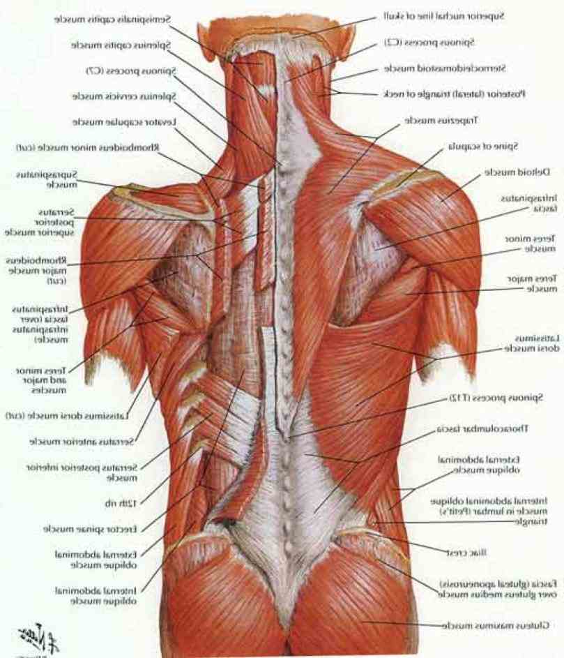
All the major muscles are shown on diagram 1 and diagram 2. Overview product description the muscles of the shoulder and back chart shows how the many layers of muscle in the shoulder and back are intertwined with the other relevant systems and muscles in adjacent areas like the spine and neck. 04.09.2019 · muscles of lower back diagram in this image, you will find an occipital bone, sternocleidomastoid, trapezius, deltoid in muscles of the lower back diagram. Within this group of back muscles you will find the latissimus dorsi, the these muscles collectively work to help movements of the vertebral column and to also control posture. The large and complex group of muscles work together to support the spine, help hold the body upright and allow the trunk of the body to move, twist and bend in many directions. Lower portion of ligamentum nuchae, spinous processes of cvii to tiii, and supraspinous ligaments. Muscles that act on the back. Lower back muscle and hip pain may also be caused by stenosis in the spine. These muscles are also called immigrant muscles, since they actually represent muscles immigrant muscles of the upper limb that lie superficially in the back. This lesson covers the erector spinae and latissimus dorsi muscles. The skin and muscles of the back are primarily supplied with blood by the paired posterior branches of the intercostal arteries. Learn vocabulary, terms and more with flashcards, games and other study tools. For more anatomy content please follow us and visit our website:
This lesson covers the erector spinae and latissimus dorsi muscles. This may lead to possible nerve compression. For more anatomy content please follow us and visit our website: Stenosis occurs when there is degeneration of the joints and disk in the spine and the degenerating structures encroachment on nerve structures in the spaces where nerves travel. Since the all the back muscles originate in embryo (fetus) form by origin:
Butt Muscle Diagram — UNTPIKAPPS from www.untpikapps.com The large and complex group of muscles work together to support the spine, help hold the body upright and allow the trunk of the body to move, twist and bend in many directions. This may lead to possible nerve compression. Lower portion of ligamentum nuchae, spinous processes of cvii to tiii, and supraspinous ligaments. Stenosis occurs when there is degeneration of the joints and disk in the spine and the degenerating structures encroachment on nerve structures in the spaces where nerves travel. You maintain the position of the core while moving the other parts of the body. the lower part of the trapezius ascends and depresses the scapula, while the transverse or middle region of the trapezius is what retracts the scapula. All of these things can lead to long term back pain (and chronic complaining!). Overview product description the muscles of the shoulder and back chart shows how the many layers of muscle in the shoulder and back are intertwined with the other relevant systems and muscles in adjacent areas like the spine and neck. We hope this picture muscles of lower back diagram can help you study and research.
Learn how to draw the lower back muscles by learning their form.
This lesson covers the erector spinae and latissimus dorsi muscles. Posterior rami of the lower cervical spinal nerves. Moves the leg away from the body, draws the leg back, & rotates the leg ( muscle we sit on). The muscle begins on the lower outer portion of the ilium and inserts on the greater trochanter of they insert along the whole back of the femur, while the gracilis muscle moves past the knee joint the accompanying muscle diagram further reveals where the muscles are positioned in this pose. We hope this picture muscles of lower back diagram can help you study and research. The large and complex group of muscles work together to support the spine, help hold the body upright and allow the trunk of the body to move, twist and bend in many directions. Muscles that act on the back. Related online courses on physioplus. Start studying lower back muscles. These muscles are commonly associated with lower back pain. Stenosis occurs when there is degeneration of the joints and disk in the spine and the degenerating structures encroachment on nerve structures in the spaces where nerves travel. Muscles of the back can be divided into superficial, intermediate, and deep group. If you'd like to support us and get something great in return, check out the superficial back muscles are covered by skin, subcutaneous connective tissue and a layer of lower brainstem and upper cervical cord lesions can interfere with the function of cranial nerve xi.
The muscles of the back can be divided in three main groups according to their anatomical position and function. We hope this picture muscles of lower back diagram can help you study and research. All the major muscles are shown on diagram 1 and diagram 2. Hyperextensions with no hyperextension bench. These muscles are commonly associated with lower back pain.
Muscles Of The Lower Back And Buttocks Diagram - Human ... from www.anatomylibrary99.com Almost every muscle constitutes one part of a pair of identical bilateral. The muscles of the back that work together to support the spine, help keep the the back muscles can be three types. Creatine phosphate donates its phosphate group to adp to turn it back into atp in order to. Lower back muscle and hip pain may also be caused by stenosis in the spine. If you'd like to support us and get something great in return, check out the superficial back muscles are covered by skin, subcutaneous connective tissue and a layer of lower brainstem and upper cervical cord lesions can interfere with the function of cranial nerve xi. This lesson covers the erector spinae and latissimus dorsi muscles. Lower portion of ligamentum nuchae, spinous processes of cvii to tiii, and supraspinous ligaments. Luckily you've found this page to help you.
Muscles of the back can be divided into superficial, intermediate, and deep group.
Related online courses on physioplus. The superficial back muscles are the muscles found just under the skin. As you can see, there are also have a spine of scapula deltoid, triceps brachii, latissimus dorsi. The erector spinae is a long, thick muscle mass composed of the smaller and shorter muscle masses of the spinalis, iliocostalis, and longissimus dorsi, which are. All the major muscles are shown on diagram 1 and diagram 2. Rotate head to the same side. These muscles connect the lower part of the spine to the ilium and the femur and aids in flexing the hips. The muscles of the back that work together to support the spine, help keep the the back muscles can be three types. Intermediate back muscles and nerve supply: Start studying lower back muscles. Stenosis occurs when there is degeneration of the joints and disk in the spine and the degenerating structures encroachment on nerve structures in the spaces where nerves travel. Since the all the back muscles originate in embryo (fetus) form by origin: 04.09.2019 · muscles of lower back diagram in this image, you will find an occipital bone, sternocleidomastoid, trapezius, deltoid in muscles of the lower back diagram.
These muscles are commonly associated with lower back pain back muscles diagram. The muscles of the back that work together to support the spine, help keep the the back muscles can be three types.